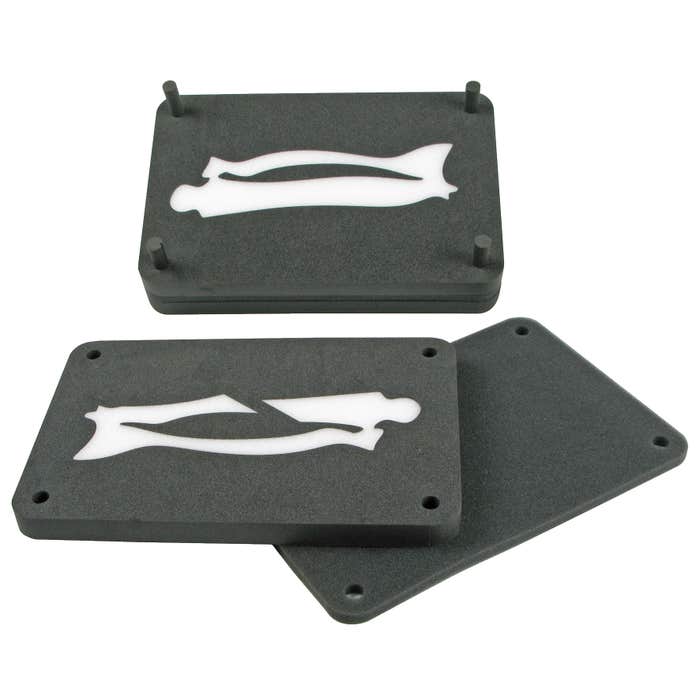
Materials: polyester foam.
Dimensions: 300 x 200 x 80 mm.
3D ultrasound model
Sku: 222060
Jeulin
The strong points
€116.45
€139.74
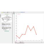
Made up of specific foams selected for their properties with respect to ultrasound, the 3D Ultrasound model conceals a healthy or broken bone. Using the ultrasound sensor, students will experimentally, through a progressive approach, establish the diagnosis by analyzing the behavior of ultrasound waves based on the sensor's position. How can we see organs invisible to the naked eye? How does an ultrasound work? The 3D Ultrasound model and sensor allow for the discovery of the principle of exploration through ultrasound, used in healthcare. Students seek to identify and interpret 2 images obtained by ultrasound (here a normal bone and a broken bone). The Scientific Workshop module provides a didactic approach to the different phases of this medical imaging technique.
Model bone normal, bone broken.
| Caractéristiques techniques |