Impact of UV on DNA Kit
-

-
Designed by Jeulin


The kit allows for the identification and quantification of DNA lesions induced by UV.
Principle:
In solution, plasmids adopt a supercoiled form.
1. The supercoiled plasmids are exposed to different doses of UV to generate DNA lesions.
2. A restriction enzyme (endonuclease) will track and cut the DNA at the site of the lesion, thereby causing the relaxation of the plasmid.
3. Separation and quantification by electrophoresis. The relaxed plasmids (linear) migrate less rapidly than the supercoiled plasmid DNA (T). The intensity of the bands (1,2,3) is proportional to the number of lesions induced by UV.
Format: 10 complete tests
Duration of the experiment: 1 session of 50 min
Storage duration: 6 months
Storage condition: - 20°C
Separation of DNA fragments: 1% agarose gel
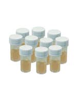
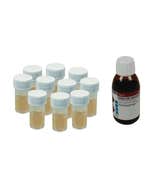
The kit consists of:
- 4 Petri dishes
- 40 microtubes
- 4 solution reagents: plasmid DNA, restriction enzyme, loading buffer, and molecular weight marker
4 Petri dishes, 1 sachet of 40 microtubes, 4 reagents in solution: plasmid DNA, repair enzyme, loading buffer and molecular weight. For 10 pairs.
| Thématiques | Mutagénèse |
-
In stock
-
In stock
- Imminent stockpiling
- In stock
-
In stock

-
In stock